November 10, 2025
Hormones and Hellfire Ep. 2: What Every Woman Needs to Know About Mammograms
Hi everyone! For our second episode of Hormones and Hellfire, we're diving deep into one of the most relevant (and sometimes anxiety-inducing) topics in women's health: mammograms and breast cancer screening.
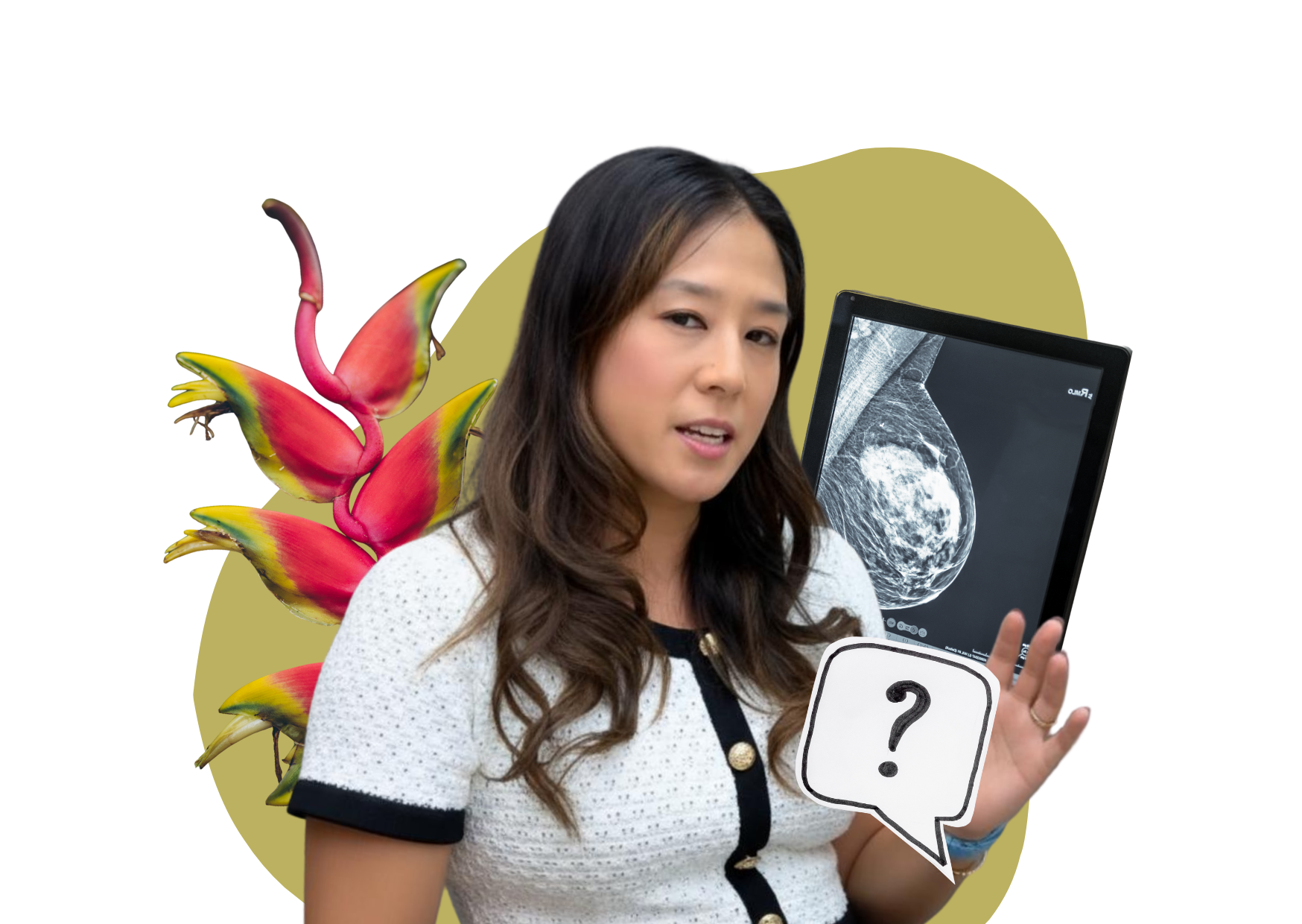
We sat down with our friend Dr. Mindy Yang, one of the top breast radiologists here on Oahu and clinical AI director for Radiology Partners. With training from the University of Pennsylvania and experience in both academics and private practice, Dr. Yang is dedicated to helping women understand exactly what happens during breast imaging and cutting through the misinformation that's everywhere on social media.
If you've ever felt confused about when to start screening, which tests you actually need, or if you should be worried about radiation… this conversation is for you.
Keep reading for a summary of the three main lessons Dr. Yang left us, or scroll until the end to find links to the full episode (and the full transcript if you prefer reading!)
Three Key Takeaways from Our Conversation
1. Mammograms and Ultrasounds Work Together — You Actually Need Both.
One of the biggest myths Dr. Yang debunked is thinking you can skip the mammogram and just get an ultrasound to avoid radiation. But the truth is, these imaging methods complement each other in crucial ways.
"Ultrasound and mammograms are almost like having two eyewitnesses to some crime scene. They have different perspectives, but you need both of them to understand the full picture."
Mammograms excel at detecting calcifications (which are often the earliest sign of breast cancer), while ultrasounds are better at identifying masses, especially in dense breast tissue. Using only one method can lead to missed cancers or unnecessary anxiety from findings that would be clearly benign with the other imaging type. The gold standard remains mammography starting at age 40, with ultrasound added as a complement when needed.
2. Getting Called Back Doesn't Mean Cancer
If there's one thing Dr. Yang wanted to make clear to all of our listeners, it’s that you don’t need to go into full panic mode if you get an abnormal mammogram callback. The statistics can actually be reassuring when you understand them.
"10 in every 100 women are called back from their mammograms. And actually, that number is higher if it's your first mammogram because you don't have priors to see if there's been a change."
Of those 10 women called back, about 6 will be told everything is normal after additional imaging. Another 2 might be placed in a "probably benign" category for short-term follow-up (meaning less than 2% cancer risk). Only about 2 out of 100 will need a biopsy — and even then, most biopsies turn out to be benign. Understanding these numbers can help you breathe easier if you receive that callback letter.
3. Thermography and Other Alternatives Aren't Evidence-Based
With so much health information (and misinformation) on social media, Dr. Yang addressed some of the alternative screening methods that patients frequently ask about — particularly thermography, which uses infrared heat mapping.
"If you just go to the FDA's page about it, you can see disclaimer after disclaimer, disclaimer, disclaimer. If you go to social media, you’ll see yay, thermography. But that's not what we (radiologists) recommend."
The bottom line? Mammography is the only breast screening tool with proven mortality benefit. While technologies like cone beam CT (which doesn't require compression) are in early stages and worth watching, they don't yet have the decades of evidence that mammograms do. Relying on unproven methods can lead to delayed diagnosis and worse outcomes.
Listen to the Full Episode
Ready to dive deeper into this conversation? We also dive deep into topics like the role of AI in radiology, what actually happens during a biopsy, and why that annoying breast compression during mammograms is actually necessary for your safety.
You can find Episode 3 on YouTube and Spotify through the links below!
Watch Episode 3 on YouTube | Listen to Episode 3 on Spotify
***
Read the Full Transcript Below
Host: Welcome to Hormones and Hellfire! We are so lucky to have Dr. Mindy Yang with us today. She is one of our breast radiologists here on Oahu. She did her residency in medical school at the University of Pennsylvania. And she's worked in academics, in private practice, and recently is now the clinical AI director for Radiology Partners.
I actually met Mindy at a women's physician group meeting. So she has a wonderful daughter and a husband who's also a radiologist, right? Okay, well, welcome, welcome!
Mindy: Thank you. Thanks for having me.
Host: Okay, so most of our, I know I shouldn't say most, our audience is like, I'd say half doctors, half just non-physicians. So we're going to go through a little bit of everything.
Mindy: Yeah, that sounds great.
Host: So today we're going to cover the basics. We've got mammograms, ultrasounds, MRIs, when you should go, how to follow up, and then all the other things that are out on social media and on YouTube.
Mindy: So maybe just to answer the question for all of our people who are not in medical personnel, What does a breast radiologist do?
Host: Sure. Well, maybe I'll start out by just talking about what a radiologist does. A lot of times people think we're the ones taking the images, but we're really the doctors who have gone through medical school, medical school training and residency and so on to interpret those images.
Mindy: So we work with our invaluable technologists, who we absolutely need to do x-rays, CTs, mammograms, which are my specialty, ultrasounds, MRI, and all of that. And then we have them pop up on the monitor. And after reviewing the images, then we'll issue a report that usually goes to a doctor or to the patient itself. And for breast radiation, I didn't actually know it was a fellowship until well into residency myself.
Host: And are most of the mammograms read by breast radiologists?
Mindy: You know, it's interesting that you say that, because as we all know, there's a major shortage of physicians going on. And that's definitely not something that radiologists are not impacted by. So we have a shortage of radiologists, but we also have a shortage of subspecialty breast radiologists. And so this is definitely a problem. You know, kind of going back to the training, we have our internship year, we then are followed by four years of radiology residency, and then a one-year fellowship in breast imaging. And most fellowships in radiology are one to two years. That being said, there are actually quite a few generalists out there, meaning that they're not subspecialized, who also help read the mammograms and breast imaging. Because without them, we just wouldn't have enough.
Host: So in a hospital system, would they have multiple different types of subspecialty radiologists? So they would have a body person and a breast person?
Mindy: And that's become a bigger trend over the last several years, decades, just because people have gotten more subspecialized, spent that extra time, and therefore they have expertise. It's the same reason why you wouldn't want an ophthalmologist doing your abdominal surgeries. Really trying to focus because the pathology is so diverse and requires so much to know.
Host: Okay, so walk us through it. As OBGYNs, we normally just do a pap smear for an annual exam, we do the breast exam, and we order the patient a mammogram. And then I send them on their way. And patients actually think that we do all the in-between because we call them with the results. So if I'm sitting in the order, the imaging center gets the order, they do the mammogram, and then you read the report and send it back. If it's normal, I just do what you have said, right?
Mindy: We hope.
Host: And then, but if it's abnormal, needing biopsies or other imaging, maybe walk the patient through who's actually leading them the way because you do that.
Mindy: Yes, so again, that's something that the general public often doesn't realize that breast radiologists are trained specifically to do. If you are called back for what they call an abnormal screening mammogram, it doesn't necessarily mean cancer, anxiety, freak out, don't do that. 10 in every 100 women are called back from their mammograms. And actually, that number is higher if it's your first mammogram because you don't have priors to see if there's been a change. And everyone's breast issue is super different.
So when you are called back, what ends up happening is a letter is issued to the patient as well because that's part of our regulations through the FDA. And that is something that we also just want our patients to understand, especially at the same age. I just feel like patients don't usually in the past have such great access to the records or understanding what that is. And we're understanding now that patients really should be sharing that decision making. They should understand what an abnormal mammogram is under the risks and the benefits of not doing the exam, doing the exam, things like that, I think are really important.
So if they are called back, then what happens is they come to our office and they get additional imaging. That can be a more mammographic view. That can be an ultrasound or it could be both or it could even be an MRI, which is typically not the first part that happens in a cycle. And all of that is performed usually at a similar facility as to the one doing screening mammograms. Once those images are worked up by a breast radiologist like myself, then we determine the next step.
There are several categories you can go. You could say, oh, it's negative and go back to screening, which is the same thing we also sometimes say benign, go back to screening. You also have a category which is really confusing called probably benign. And that is something where we put patients into a follow-up. So it's not just that we say, oh, maybe it is, maybe it isn't. There's actually statistics behind this. There's science behind this for why we do this. And anything that's put into a probably benign category has less than 2% malignancy risk. And so essentially what happens is we follow those patients at a closer interval to make sure those findings we're saying are probably benign, don't change or resolve. And that can be for a number of things.
So there is unfortunately a high number of people who get into that category. And sometimes it feels like you're kind of in limbo, just hanging out. But patients typically should understand that actually the risk of anything being super suspicious is quite low.
Host: Yeah, you're just being extra careful.
Mindy: Exactly. Now, on the other hand, if we do see something suspicious and that would be anything from 2% probability malignancy and up. So it's a huge range again. So there are certain things that we don't think are that suspicious, but we want to make sure. Fibroadenoma, very common. I'm sure you've seen that in your reports, in your pathologies. And we often biopsy those just to make sure that's what it is. Because sometimes cancer and benign things can look similar and have overlap.
So if we do recommend a biopsy, we usually tell the patient at the time of the diagnostic evaluation. If I'm working remotely, I'll call them or video conference them to talk about that. If we're in person, which is the more traditional setup, then we go in and talk to the patient about that as well. Either way, we're expressing to the patient, look, there's something that we think is slightly concerning. We want to make sure there's nothing atypical growing there. And so it's really important to sample it. Then we are the ones who then do the ultrasound-guided biopsy or the mammogram-guided biopsy. And people also don't always know that, but as breast radiologists, we do quite a few interventions.
Host: I also didn't know you could do a mammogram-guided biopsy.
Mindy: Yeah, so that's called... The fancy word for that is a stereotactic-guided biopsy. So I try to...
Host: Yeah, exactly. Sometimes we don't know.
Mindy: But things like... I also will get into this later, I know, but the reason is because there are certain findings we do not see well on the ultrasound, but we see really well on the mammogram. And so patients don't really realize that, but sometimes that's the case. That's why we need both of them together. And so what ends up happening is... The procedures are all very similar, but the patient then comes back to us and we have to sample them. But we are the ones who do it.
Host: Awesome, interesting. Okay, so in that context, then tell me the bi-rads. What number do I want?
Mindy: Oh, okay. So the number you want is a one or a two. One is basically what we call negative, okay? Two is benign. Both of those things are normal. People just have learned in different ways how they've been trained to report things. For example, you might have calcifications. You can have a huge number of reasons why you have calcifications in your breasts. The majority of which are actually benign, non-cancerous. And some people like to report that and say, oh, okay, that's benign, so I'll put it a two. Others will call it a one, even though they're seeing the same thing. It's just a difference in how the breast radiologists preferentially report things.
Host: So for the biopsy, so when you're talking to the patient, I assume in their minds, they're thinking, oh my gosh, it's a surgical procedure. But they kind of get that, why would they go get that image, right? So if they're being called back, it's that same day, or if they call back...
Mindy: So biopsies, generally, we try to schedule them not on the same day. Although some facilities will do it. And part of that is that we often just don't have enough staffing to support that. And the second part is orders, insurance can get very complicated. You may need certain insurances to clear it, and you may not be able to do that on the same day. So it can get quite complicated. And most facilities, typically nowadays, will try to schedule it on a separate day. Although it shouldn't be months down the road. We try to get patients in as soon as we can, depending on what's available.
Host: Because that's usually what we get, is the call panic, right? It's abnormal, they're telling you to call back, why can't I get in today, tomorrow? They want an answer right now.
Mindy: And we're like, well, usually they want to call you back. But essentially, all the biopsies are needles, right? They're needles that are stuck into the tissue. You're taking a sample from that, and then you send it off. The results come back in two or three days. And yeah, I give a little bit more leeway than that, depending on how bad the pathologist can be. But in terms of the biopsy, I always tell my patients, it's not a big surgery. It's not. And that helps to kind of calm down the nerves about that. In fact, we typically don't even have stitches or anything like that.
Essentially, what I tell them is, you come in, similar to how you might even get that ultrasound, if you're getting an ultrasound-guided biopsy, you're going to be laying down similar to that. We clean off the skin, and then we give some local numbing medication in that area, which is lidocaine, which is really similar, right? If you've ever had a tooth pulled or any dental work done, same thing. So we numb up that area. You don't have to be sedated. You don't have to have someone pick you up. Although, of course, we do get the occasional patient who says, why can't I have that? But I usually tell them, hey, you're going to get that pinch and burn, that sting that you get from any kind of numbing. But after that, you really shouldn't be feeling sharp pain.
I do tell them you might feel tugging, pulling, that kind of thing, because sometimes they're not used to that. Or you might hear a loud noise. I like to actually show them what it's going to sound like when you do take that biopsy sample. Sometimes it sounds a little bit more intimidating if you don't know what to expect, right? So all those things, once we go in and take a few samples, send them to the lab, I tell them, let me put a bandage over you. Usually you don't need any kind of stitches and you don't really have many kinds of restrictions. We just say, don't lift too heavy and you might get some bruising. That's normal. It might be a little sore later on when the numbing wears off. But those kinds of expectations help to kind of understand it while you're not getting put under for a big procedure. So the results come back.
Host: Do they come to you or do they come to me? Because usually they come to us.
Mindy: Yeah, both is the answer. There are certain practices that will have the radiologist call when they have results. They will personally call the patient. Other places have nurse navigators in charge or there are places that will call your office as well. So it really depends and varies.
Host: Okay, what is the difference between mammograms and MRIs and ultrasounds?
Mindy: Okay, that's a great question. So mammograms are basically x-rays of your breasts, right? There's a 2D mammogram, which is essentially you end up with four images. There is a 3D mammogram, which is now really the standard of care. And those are basically taking really thin, low-dose x-rays at different angles of your breasts. And it's like almost like book pages. So you can flip through those images. And the reason that's standard of care now is because they determine basically that we can see more subtle cancers earlier on in all sorts of breast density. So we can see them in really fatty breasts all the way to more dense breast tissue. And so that's that.
That one does have radiation. It is a low, tiny dose of radiation. Radiation is always a concern of patients, of course. But I don't know if people know this, but you're getting radiation in your background as you walk around every day. And if you're at higher altitudes, you're getting more radiation, right? That's just part of nature. And sometimes I think people don't know that. When you take a flight cross-country, you're also getting a little bit of radiation from that flight. So a mammogram basically has the same amount of radiation as taking that cross-country flight for about seven to eight weeks worth of natural background radiation.
And it's actually one of the lower dose radiation exams of all the radiology exams that we have. And so I think there is definitely some fear-mongering and misinformation out there spreading this concern about over-radiation from mammograms.
Host: Well, I think the other thing that I get brought up is that you have to do it every year. So it's the total, starting at 40, let's just say, for those who need it earlier. I still think over time, I think over the next...
Mindy: When do we stop mammograms? We typically don't. And we don't recommend that from a radiologist. Some people might in certain guidelines, but I'll tell you also on that note is that I've biopsied a very with it 94-year-old woman who has, she has the capacity to deal with treatment and to go through that and have great quality of life and better quality of life than if she didn't do that. So we kind of say, as long as you're gonna proceed with treatment, then keep screening.
Host: So the long-term exposure, do you have a comment on that?
Mindy: Yeah, it's a huge... Yeah, so our myth buster is basically that there is way lower risk of inducing cancer from a mammogram than the risk of essentially getting breast cancer. So I think that, again, risk-benefit. You know, I'd rather catch my cancer and have a minimal dose of radiation. And this is something that we've studied for a long time. And also mammography has been proven to save lives. And it has been proven specifically to save lives when you start at 40.
Host: And mammograms versus MRIs versus ultrasounds.
Mindy: Yes, back to that. For all of those. Yes, so mammograms are the ones that have radiation. As we said, it's a very low dose radiation, but it is the standard of care. And it's the only proven tool that has shown us to drop mortality for breast cancer screening. 30 to 40% is the mortality drop of having breast cancer screening.
Ultrasound is used as a compliment. So I like to say that ultrasound and mammograms are almost like having... The best analogy I can think of is two eyewitnesses to some crime scene. They have different perspectives, but you need both of them to understand the full picture.
Host: That's a great description.
Mindy: You know, because sometimes people will say, oh, it's like you can see one thing and the ultrasound shows you everything. It's just not actually true because ultrasound can't see very well, for example, calcifications, which are a big part of how we detect early breast cancer. So it's really important to have the two things hand in hand. Additionally, mammograms, conversely, especially in dense breast tissue, don't always seem the best because it's almost... We like to use the analogy of dense breast tissue, a breast cancer can be almost like a snowball. You're searching for that snowball in a field of snow. And so you can have certain masses that are obscured and having ultrasound helps you to get more information about all those things underlying.
Host: So is the dense breast... I feel like this comes up in all screening guidelines. Now, is that something that is a clinical diagnosis or is it a radiology diagnosis? That's a great question.
Mindy: It's a radiology diagnosis about... When we talk about breast density, we're talking about the amount of fat versus glandular tissue that you have. And we have four categories, essentially. We go from almost fatty, scattered, fibroglandular tissue. Then we go to heterogeneously dense and extremely dense. 40 to 50% of women fall into the latter two categories. So it's actually a pretty high proportion of the population. And with that, breast density, the denser your breasts are, it's actually an independent risk factor for increased breast cancer. And the second part is what we were just talking about, is that with denser breast tissue, mammograms are not as sensitive. So we often recommend, depending on your risk level, supplemental screening, such as ultrasound or MRI.
Host: Okay, so let's speak of the MRI. So we have a lot of high-risk breast cancer patients. We alternate mammograms plus MRI alternating six months. A lot of patients will ask me, why can't I just do the MRI every six months? Why do they do both modalities and why is it six months?
Mindy: So the proven benefit really still lies in the mammogram, right, as we were talking about. Even for an MRI, which is a highly sensitive test, and why we use it in high-risk patients, there's a few reasons why it's not the only screening tool we use. First of all, it doesn't see calcification super well. Sometimes you may have early breast cancer that doesn't enhance super well, that you might see on the mammogram, but you don't see as well from the MRI. Other reasons are cost and also proven, right? A proven benefit of screening mammograms on mortality has been shown. But the same is not true for MRI. MRI does not necessarily show an actual outcome benefit when you look at mortality.
Host: Is it mainly because of the calcifications?
Mindy: Well, there's a few different reasons for that. That is one of them. But again, the other thing with MRI is that you have to keep in mind, because it's so sensitive, it is not actually as specific. And so what ends up happening is you have a lot of findings and you might end up therefore doing more biopsies and becoming more invasive. It's a little bit of Pandora's box sometimes. And so that's why we say, it's kind of on the patient's understanding that they are okay with doing more invasive procedures if it means catching that extra cancer, right? So all of these supplemental screening methods, the reason we use them is to detect additional cancers on top of what we would do just with mammograms. And they do show detection of a few extra cancers, but at the expense of having more false positives, more biopsies.
Host: I feel like I am familiar with the, in my practice I was doing the mammograms with the MRIs. And I mean, that's what I get because I'm at higher risk. So the question I had is that in California, we were at a thing recently with a bunch of lawyers and they all talk about how they do mammograms with ultrasounds, like kind of alternating. Is that like location dependent or is that just?
Mindy: Well, there are regional patterns, but part of it is also looking at risks. So our American College of Radiology has recommended that beginning at age 25, you have a risk assessment. That takes into account things like your family history, hormonal therapy usage, whether you've been genetically tested for mutations like BRCA1, BRCA2. And then once you have that risk, you can kind of then plan better. That may change in the future as we look at some of the things that are coming out with AI and all those things in terms of risk. But right now what we recommend is anyone who has a calculated lifetime risk of over 20% falls into that category where we call them high risk. And high risk, therefore, then we do recommend, generally speaking, mammogram and MRI, usually alternating every six months. But some of the practices do differ.
Now, if you're under that, that's where you're kind of in a gray zone of, should I get supplemental screening or not? And a lot of the time what we do right now is we may recommend you consider supplemental screening. Like if you have dense breast tissue, if you're in those two categories that I said, you might wanna get ultrasounds. But I do think it's somewhat of a personal preference in terms of patients understanding what is your density? By the way, to do that and figure out what your density is, you have to look at the actual report. The other thing is that is now sometimes being included in patient letters because of a law that was passed last year. So it should be in both. But if you have any questions about your breast density, look at your letters, look at your past reports.
But once you determine that, are you the kind of patient who's okay with getting potentially more false positives? Would you have peace of mind at least having the option to biopsy something, even if it's more likely to be benign than not? So I think everyone has a different kind of tolerance for that. So I think that that's only now kind of becoming understood more.
Host: Well, that dives into the, okay, the fear mongering. We have so many patients who decline mammograms, more so than we did five years ago, 10 years ago. And some of it is, I hear about it as painful. The procedure is like, why do they have to, is there any other way than squishing my breast?
Mindy: Yeah, that's a big one.
Host: And then the radiation exposure.
Mindy: Yeah, well, the first thing I usually say is that clearly a man invented the mammogram.
Host: Oh my gosh, 100%, I say that all the time.
Mindy: I cannot deny it. It's not comfortable, right? I've gone through it myself. I know it's not comfortable. But I also think a lot of it is your expectation. I think if you understand why we're doing what we're doing, then it makes more sense, right? So breaking it down is the reason that we need that compression to be a few fold. One is it actually reduces the radiation you get to your breast. Two, it actually makes the images way more crisp and clear, which is really important, especially when it comes to calcifications, because you'd be surprised by just a little bit of motion, how hard it is to determine whether calcification is benign or malignant sometimes. So the compression is actually super important.
And finally, it's a very temporary kind of pain, I think. There are many tests we do out there that have a little bit of pain. You might say that for when we get flu shots, that's a quick pinch. It's not that different to me. I think it's just pap smears.
Host: Pap smears, I personally think a pap smear might, for me, but where's the email of a scam? But people do them, right?
Mindy: And I think a lot of it is just because of the media. I think media and culture and not having enough people go out there and tell their experiences, because a lot of times it's also, to me, patient preparation. If they know more about the test going in, which is why we're doing these things, right? Talking about it. If they know more, there's less fear. It's even how I can tell in a conversation with a patient, if they're freaking out about a biopsy, and then I kind of say like, okay, but it's not a big surgery. You don't even have a band-aid. They quickly calm down. And I think it takes just words. And as we're getting into more and more of a, I guess, certain influences by social media, influencers, things like that, I think it's important to have people speaking out about their stories, because most patients will tell me, well, that wasn't as bad as I thought it was going to be. And so for me, it's an expectation.
Host: Well, there's this one I was going on the deep end doing research on where you lay on a table and it doesn't squish your breast, but you mentioned that it lessens the radiation with that squeeze. A common comment in Reddit was that it intensifies it by squeezing it because you're now just beaming the x-ray beam to the breast. I don't think that's how it works.
Mindy: That's not how it works. There's a reason we have physics on our boards. And physics is, I'll tell you, I'm super bored by physics all the time, but there's a reason we learn about it. There's huge sections on breast imaging physics for this reason alone. It's counterintuitive. Some of the things are very counterintuitive. For example, we've talked in the past about shielding, right? And a lot of shielding now is no longer recommended by radiologists. If you look at, again, if you have any doubts, you can always go to the American College of Radiology. They have the most up-to-date guidelines. And if you're a patient, you can go to radiologyinfo.org, which also helps explain a lot of different exams in patient-friendly language. But shielding actually counterintuitively can cause increased scour because of the way physics works. So it actually can counterintuitively increase your dose. But if you're not a physicist, why would you know that? If you're just a layperson, why would you know that? Of course it sounds better to have a shield. You don't know, right?
Host: I have doctors asking me these questions all the time too. So it's a very nuanced subject. Right. My only thesis is that what goes in must come down.
Mindy: That's a good one. But going back to your question also, I've heard of that. It's actually a newer technology. So we have cones. It's kind of a CT of the breast is what has been done. And it is touted as something that doesn't really require compression, which is very appealing to a lot of people. It's in the early stages. So that one is not, it's not something that I would just blow off, but it is something that is still in the early stages. And so remember, we have many studies actually showing proven mortality benefits of mammograms. That is our gold standard. We know it works. And that is the best we have right now. Until we have that for other technologies, then I wouldn't be able to say that I would recommend one without, on its own.
Host: Well, with that comes the fear that most patients are saying, hey, I just want the ultrasound. There's no radiation with it. They're supposedly more comfortable, which I feel like it might not, because that thing is, that part is pressed really hard. You remember it like an echo or anything, you've seen it, or we do scanner things. It's really hard, right? So the ultrasound, so that's our number one thing that I would say right now is that everybody wants it. We get a lot of patients who decline the mammogram and just want the ultrasound.
Mindy: And I think it really comes back to the fact that they're complementary, right? So one, mammograms, as I said, is the gold standard. But back to the analogy of it is, there are two different eyewitnesses giving you information, different information about your breast tissue. Both are important, but you need both. For example, you could find a really suspicious finding on ultrasound. And if you don't have a mammogram to correlate, you can start going down the rabbit hole of, oh, then we should biopsy this. Only using ultrasound is ultimately, of course, going to lead to more biopsies.
A really good example of that is if you get bruised from trauma, or if you had a fall, motor vehicle collision, we see this not infrequently, you get bruised, we see a pretty ugly looking thing under ultrasound. It often looks almost like cancer. But if you know the clinical context and you say, okay, I need to go get a mammogram. So you have the mammogram to correlate, you can see fat necrosis. You can see the trauma very obviously on the mammogram. So that's an example where if you didn't have a mammogram, you'd be stuck.
So even though I know it sounds great, and to your point also, people often think it's going to be really comfortable. You're not, unfortunately, at a spa. You're just not. I would like it to be. And of course, there are many ways we can make it more comfortable from the patient experience. But I think the problem with ultrasound alone is that it's also very operator dependent. You can find that there are, I've seen so many cases where people decline mammograms and there are random shadows, random masses that just aren't real. And that's causing a lot of anxiety in patients who ultimately then essentially repeat the whole evaluation. And so it's just almost a waste of time, which can lead to delayed care, delayed diagnosis of breast cancer and bad outcomes.
Host: Tell me about thermography. Thermography. This is, I feel like the only thing I hear tons about and I'm like, no.
Mindy: Yeah. So if you just go to the FDA's page about it, you can see disclaimer, disclaimer, disclaimer, disclaimer. If you go to social media, you can say, yay, thermography. But that's not what we recommend. There's a reason we don't recommend it. So yes, it's essentially like an infrared heat map. Right. And they kind of tout it as something that can show potentially breast cancer earlier than you even see physical changes.
It's not a radiology supported modality. Not for anything. I think that these kinds of companies that tout this have to come out with a disclaimer saying this is not supposed to be used alone. So if you ever see anyone saying this can be used in place of mammograms, that is actually against FDA rules and they should not even be saying that at all. So that's misinformation. But we really have seen quite a few patients come in with abnormal thermogram findings, very anxious. Again, they go through all this repeat diagnostic stuff that they could have just done to begin with to avoid any of this. And there's nothing there. Or the opposite where they rely completely on the thermogram, which doesn't necessarily show anything until it's too late. Because our gold standard, the mammogram, shows us the earliest signs of breast cancer. So if you rely on some of these modalities that are not evidence-based, then you run the risk of coming to us later with a poor prognosis and worse outcome.
Host: Who is actually reading these scans?
Mindy: So these scans are generally not read by any radiologist. There are certain professions out there that will read these. But, and I respect the fact that all professions should be respected. Everyone has a right to decide what they do with their body. I think that's very much something that I advocate for, for patients. But they should also understand what scientists and radiologists who have been spending their years really studying these things and learning about this recommend.
Host: Well, what was I going to say about the bar? Well, you know, I was reading, I saw a YouTube commercial about it. It's very convincing, right? Because it says, hey, the cells speed up when they are multiplying, they heat up. And I was like, well, that's some basis in science there. And then they lead you down this great way of like how easy it is to detect and how much sooner than it is for calcifications, because calcifications take a while for breast cancer to lay down. I'm like, wow, I could see somebody who doesn't understand the medical part of it or... It's just not science.
Mindy: Yeah, I mean, I think that speaks to the culture that we're in right now that we as physicians do have a duty to speak up against, because I think that there's a lot of superficial knowledge out there. And so therefore, people who don't necessarily know that much are attributing factual claims to opinions. And so I am concerned. I do think that we see a lot of patients who are rightfully confused. And so I do think it's important for us to take the time and focus more on that. And even with patient reports in radiology, that's something that's actually very important to me, because I get reports from friends and family, including physician colleagues, all the time, because they don't understand them. Or I'm sure you've heard, oh, what? Is my chest radiograph unremarkable? Like, why is it not remarkable?
So the language we choose to use in our reports, which in breast imaging is very structured, is something that I think we should consider in the future about how we address that with patients and having a better patient-centric experience, patient-friendly reports, which people are starting to do and understand. But we're still behind, you know.
There is actually a great website called Cancer Besties, which you may have heard of, but you can insert, and I'm not affiliated with them at all. It's just something that I actually really like for patients. And it's made up of actually non-doctors, and it's a breast cancer survivor, and a pre-vivor, somebody who had a preventative mastectomy is done for risk, and you can drop in your report, and they explain it in patient-friendly language, and also talk about what questions you can ask. And I've tried it a couple times just to kind of vet it out, and it's pretty good.
Host: Do they use AI for that?
Mindy: They do. I believe they do. It's not explained, but I would assume that they're using some sort of generative AI for that, yeah.
Host: Well, that leads us to the next slide. Well, that's another thing, too, is that on Reddit recently, they think that radiologists are using AI and reading all their images. Can you comment on that? Because this was a comment for somebody who said, well, I'd rather go to a non-physician who gets one-on-one care for me, and read the images, rather than an AI program. And I was like, I don't, that is not even, AI is nowhere close to that. No, no.
Mindy: And I think that there are a lot of sensational headlines out there, because everyone wants to talk about AI replacing doctors, or any other entity really, now and then, right? But AI, we really consider it's augmenting us right now. It's a tool that, as you said, it's not perfect. And it's interesting, because I was having a discussion with another physician friend the other day about how we expect, our expectations for AI is different than our expectations for humans, right? So we expect AI to be perfect 100% of the time. It is definitely nowhere close. So what is acceptable? That hasn't been determined yet, right?
So looking at breast AI specifically, how would I feel about AI being the only sole reader of my study, but not being perfect? Is that better or worse than just having a human reader? Usually the answer is neither alone is perfect. You need humans plus AI to be better. And we're still working on improving them with foundation models and all sorts of kinds of multimodality models coming out. I think we're starting to see the future is going to be bright, but we have a lot of obstacles to get through to get there.
Host: So you're saying if I had an image of a mammogram, the human plus AI is more accurate than the human alone and AI alone.
Mindy: Correct. In most models, that is currently true. Even though you will see certain people saying, oh, well, they performed just as well. There are often many caveats and nuances to that. And many of the studies you have to look at in different contexts, because, for example, in Europe, a lot of the countries there do double readers for mammograms as their baseline.
Host: So the reader is getting like two positions where it really works itself.
Mindy: Yeah. So it's very common in Europe compared to the U.S. and, of course, different medical legal contexts. And so there's a lot of contributing factors to what we're seeing.
Host: Okay. And then what else is AI being used other than the reader mammogram? Right.
Mindy: So this is where I think it gets really exciting. In the past, as a breast radiologist, we always kind of poo-pooed old AI because you may know about computer-assisted detection CAD. And we were like, oh, it marks up like everything on the breast, so it's not helpful at all, right? All these false positives, essentially. And now we're seeing AI not just in diagnosis, not just in detection. Where we're seeing it, which actually is exciting, even though it sounds boring, is taking care of all the boring crap. Like, I don't want to sit there and have to go tell the technologist, oh, you didn't get quite enough muscle here, so let's bring the patient back and torment them after the patient's already left. How about if the AI program tells the technologist before they're even done with the patient, hey, you got to get a little bit more tissue here. That's a great use of their time and not a good use of my time, right? And then things like tasks that we have in report creation, kind of going from the image to the report. So things like that and automating a lot of those administrative tasks or what we call in radiology non-interpretative tasks has become very burdensome or akin to paperwork, right? And so we're trying to lessen that burden so we can get back to just focusing on interpretation.
Host: No, I think that goes to say exactly what you brought up earlier, is that there's less physicians so that we need to maximize your time.
Mindy: Yeah, we talk about improving capacity a lot of radiologists and we're all burned out, right? Like, we all know it. Like, I feel like burnout is a word that you don't even want to hear anymore because it's just every day. But how do we lessen that and still provide high-quality care to the patients, right?
Host: Tell us more about your AI projects.
Mindy: So I'm really excited about all the different parts of it. I have interest in everything from what we're talking about with report creation and non-administrative tasks for radiologists. Of course, I'm always interested in detection. You want to improve those models. But I also am really interested in patient-centric things. So patient-centric reporting or even in the past we used to be known as almost like doctor's doctors, right? We would be like a consultant for the doctor where we'd have all this time spending talking to referring teams. They would come down from the waiting room.
Host: We were doing this all the time in residency.
Mindy: And it doesn't happen anymore because no one has time. Nobody has time and we're all burned out. So how can we kind of get back into that mode where we're all operating as more of a team, including the patient, where the patient is in the middle. The patient and the family are in the middle. So we always forget about caretakers too when it comes to oncology. I feel like that's such a big component to the team. Just even walking them through what to expect from coming into the department, how you're going to be waiting for your mammogram. They should be able to have all that information, what a biopsy feels like. And kind of just improving that experience for them, I think, is really vital. You know, back when, it's not even that long ago when I was in residency, we didn't have breast care teams.
Host: No.
Mindy: You got the mammogram and then goodbye. And then it goes abnormal. Here's the oncology. But now, most places have these really built out teams with multiple layers of where you can go that help patients go down that journey. Well, another thing that is really exciting is things are getting more personalized, right? We're learning more about tumor receptors and personalized treatment. And even from the images themselves, this is exciting to many and scary to others, but you can find information in the pixels that we as humans cannot know and that are predictors for potential risk. And so I just feel like there's so much opportunity potentially for really being able to hone in on those truly high risk patients and the patients who may not fall into our typical traditional high risk, but maybe the images combined with their contextual history and things like that would offer us something otherwise. So I think we're looking outside the box and when we start doing that, understanding then how to guide more personalized treatment as well.
Host: No, I think that's a really nice compliment for all these people that are looking into longevity medicine. It's more than supplements, it's all of this stuff. Knowing that you are at a higher risk and then kind of acting on that and maximizing your early detection for these conditions.
Mindy: Most breast cancers are in people who do not have family histories. The first thing people want to say is, well, but I don't have anyone in my family who has breast cancer. That doesn't mean that you won't get breast cancer. Unfortunately, as we know, one in eight women. And then if you're looking at ages 40 to 49, it's actually one in six. And we've seen that the younger women have the more aggressive breast cancers. And so we're really trying to figure out how to intervene at an earlier stage so that we aren't seeing those numbers.
We have been working on several AI projects, and the thing you mentioned about the pixel is that AI doesn't see the world that we do. I think that a lot of people, we didn't know that until we got into it deeper, where you put the information in and it may find something that we have never seen, and it recognizes continued pattern as saying this is a high risk versus low risk, something that you and I just don't even see. That's the cool thing about it. And we also don't know how it knows that either.
So sometimes that black box transparency issue is, of course, an issue that we're still grappling with and how to explain that to the general public even. How much trust do we have in AI as physicians? And it's quite a different scale depending on who you talk to. But then also we want our patients to trust us and to trust AI, but not over-relieve, not over-trust. So where's the kind of balance, right?
Host: No, there isn't.
Mindy: There isn't right now.
Host: Not yet.
Mindy: That's why it's a wild, wild west.
Host: Yes, 100%. Okay, we did answer the question, do breast mammograms prevent breast cancer?
Mindy: Yes, they certainly — oh, wait, sorry. They do not prevent breast cancer. But they detect breast cancer.
Host: Yes, earlier, sooner, gives you the best, and you talked about mortality, that means the ability to survive breast cancer.
Mindy: Exactly, exactly.
Host: We're specifically a perimenopause, menopause company. Do you have any comment on how you treat patients like that differently on imaging? Does it look different? What do women at 40 to 50 see versus someone who gets early mammograms?
Mindy: Yeah, that's a really good question and we do not treat them differently, but the images often do change over time and that is because your breast density tends to change with hormonal fluctuations, right? So as you progress, perimenopausal and postmenopausal, you generally have increased fat and decreased glandular elements in your breast tissue. So if anything...
Host: It is sad, I know.
Mindy: It's sad, right? That's why I said that. The funny thing is, as a breast radiologist, it's great for us because it actually makes the mammogram easier.
Host: Yes.
Mindy: So I'll sometimes see the person come in who's been faithfully coming for 5 to 10 years and I'm just like, oh, okay. Like now I can see that area much better. Yeah. It's less fuzzy right there. No, it's not as smudge on my screen. And the other thing is, a lot of patients also, the reason we ask so many seemingly invasive questions before you get your mammogram is because things like hormonal therapy, OCPs, birth control, that can affect what we expect to see on your mammogram and your ultrasound as well. And so it's important for us to know. We're not just trying to be nosy. Actually, some patterns that I do not see infrequently are patients who go on hormonal replacement therapy. What I'll see is they actually have increased glandular tissue, right? In certain areas of their breasts. And because of that, they might get called back because there's that change. And so we look at it, but I've seen it enough to know, okay, that's probably because they're on hormonal therapy.
Host: We start screening at 40. Every year until whenever you won't get treatment, which I think is a really nice way to think of it. Because if you're not going to get treatment, then there's nothing.
Mindy: I used to think of it as like longevity. Like a standard of… what word am I looking for?
Host: There's a brain fog.
Mindy: The brain fog is hitting. Life expectancy? QELs! I was so close.
Host: Well, they also use life years gain.
Mindy: But that's like a society breast injury thing that they say. But I don't know who else uses that, but I like to think of it that way.
Host: So explain that for me. What is that?
Mindy: Life years gained. Like basically, in your screening marker, beginning at age 40, saves the most lives, but also increases life years gained.
Host: I like that.
Mindy: Yeah. So people kind of have an idea. For every 100 examinations, 90 of those women will be told that it's normal or benign. And then 10 of those will be called back. As I said earlier, baseline's a little bit higher. Of the 10 that are called back, about six will be told at that point that it's normal, negative, or benign. And then two of those may go into that category of probably benign when we follow them at those six-month intervals up to two years. And then two of the remainder will essentially be recommended to have a biopsy. But also, I want to say by and large, most biopsies are benign. And so there are multiple different categories that you can kind of dive deeper into in terms of the risk of malignancy where we talk about a specific lesion having a higher malignancy rate than another. But I don't really think it's always necessary because a biopsy is a biopsy.
I do tell my patients if it's something, what I call, BioEdge 5. So that's when you have 95% or greater probability of it being malignant. We talk about masses and how their shape is, where they're located, what their margins are. And our margins are like how you kind of can go around the outside. And if it's something really suspicious, such as what we call speculated mass, and that's kind of like a spidery looking mass that's really ugly, then I will tell my patients, look, I'm really concerned about this area. I do think it could be breast cancer. Tissue sampling is the most important thing for us to do at this stage. I don't want to sugarcoat it even though I obviously take into account how the patient's responding to things because I want them to understand the importance also of what we're doing.
Host: What's the difference between a screening mammogram and a diagnostic mammogram?
Mindy: Perfect question. So, screening mammograms are done for those who are asymptomatic, meaning they don't have any concerns. Anything outside of that you can be called back for, sorry, anything outside of that is a diagnostic evaluation. So, a diagnostic evaluation, diagnostic mammogram can be because you're called back for a screening mammogram or if you have a lump or if you have pain, you have nipple discharge. And this is actually something I would love to discuss with you, suspicious nipple discharge. Sometimes you also probably see this. We tend to say if it's unilateral, bloody, those are super suspicious. Unilateral, of course, if it's blood tinged or if it's clear even sometimes you can get clear nipple discharge, which you can have for a lot of reasons. But if you have something more oily, gunky, that's usually physiologic. If it's bilateral, we usually think it's kind of just something that your body's unfortunately producing.
Host: Yeah, yeah, yeah. I always think bilateral is okay. I'm less worried.
Mindy: Unilateral, okay. And we were meaning one breast versus two breasts.
Host: I get a lot of questions about nipple discharge at parties.
Mindy: At parties?
Host: Yeah, they'll be like, oh, by the way, I have nipple discharge.
Mindy: That is what we say all the time, too. We say it to people. I always tell my patients, I'm like, okay, so your nipple discharge is probably fine, but don't keep touching me. Just stop. I know you're curious, but stop. It's going to get worse. Yeah, so, and then the other thing we like to say is diagnostic mammograms. So those are different views than the screening mammograms. So screening, you get your four standard kinds of positions. With diagnostics, if you're called back for certain findings, then what we do are certain views. For example, for calcifications, you get what are called magnification views. It's literally like taking a magnifying glass. So it really depends on what you're called back for.
Another thing is that because we have such structured language, patients tend to freak out a little bit about the language we use because we use words like mass, distortion, asymmetry, calcifications, and that means a lot of different things to different people, especially mass. So I just want to tell patients not to get scared of those words. Those are something that have been studied and standardized over time, and it was partially because we didn't, at that time, expect patients to be reading their reports. And I think now we're starting to adjust to understand that we need to be more mindful of that. But a mass can also just be a cyst. You know, so a cyst is a fluid-filled collection that is...
Host: Oh, a fluid.
Mindy: Exactly. That most of the time is not something we need to be concerned about.
Host: We love when radiologists call us and correct the order that we have put in. I don't know how many times...
Mindy: I have no problem with that.
Host: I have no problem, yeah. Sometimes we're in a hurry. Sometimes it's not the right thing.
Mindy: I don't... I straight up admit that occasionally I just don't know what to order.
Host: But that's great because honestly, that's great as a radiologist because I feel like we should be the experts, right?
Mindy: Right.
Host: We're the ones who are asking for it and we know that. But for a good reason, we can't self-refer. We can't say, go do this.
Mindy: Right.
Host: Because obviously there's a conflict of interest there. But there's a reason that there are things like flex testing, right? Like if you put in, if needed, right? And I think people get confused because we spend a lot of time, our front staff spends so much time on the phone trying to talk to doctors about why we need both sides or if we're only having pain on one side, why do we need another? Well, you'd be surprised how many cancers you incidentally find on somebody who only has symptoms on one side. And everything we were just saying, right? You want to compare. You want to compare symmetry. You want to compare things like that. Sometimes we need the ultrasound, sometimes we don't. And I think it can get frustrating from a scheduling standpoint, from a referring standpoint because you have these slots that you don't know if they're going to be used but it allows us to do what we need to do.
Host: Right, right. I don't know. I don't know what physicians argue with you.
Mindy: I know. All right. All the time.
Host: All the time. Yeah.
Mindy: I will do it.
Host: Okay. So to wrap up this episode, we have a few more, we have a couple more questions for you. First, what's the one thing you wish someone had told you about breast cancer prevention?
Mindy: I think that people underestimate how much they can control their own prevention. Right? So exercise and weight maintenance but also alcohol intake and even understanding how your birth control and those kinds of things contribute, just understanding risk in general. I actually probably like up to three or four times a week pasting the link to the online calculator for calculating your own risk. If you search to find your risk through MagView actually, you can plug in your family history, your when you started your periods, if you had a baby, all those things people don't necessarily realize it really contributes to your overall lifetime risk and your five-year risk and things like that. So I think understanding your risk status and getting more familiar with that so that you know what options there are is something that I wish I had known.
Host: And our final question is share one of your current obsessions that you have right now. Books, podcasts, videos, TV shows. I'm obsessed with TV shows.
Mindy: So for some reason because everything I think about these days is probably with my obsessive toddler. I wouldn't say it's an obsession, but I do have a very soft spot for the TV show Bluey.
Host: Oh my gosh. Oh, Bluey's great.
Mindy: I love it. Yeah. It's so good. Right. Exactly. And I feel like it's so funny because I was always like, ugh, I'm not going to be that mom who loves all the kids shows. But now I'm like, Bluey's great.
Host: Bluey's so good.
Mindy: She doesn't even waft to you that often, but I just think about the fact that like, it's such a great, I have so many friends who are like, Bluey made me cry because I was talking about like working as a work mom and also just, you know, imperfect perfection, right? So I don't know. Something about that just really makes me cry.
Host: It's a working mom.
Mindy: No, it is. I love it. It's like, things that are twisted, I think we all need a little bit of levity right now. So things like that are, I find oddly a lot of joy in that.
Host: Thank you so much for joining us, Mindy!.
Mindy: Thank you.


Real Talk, No Filter
Join our newsletter, where we talk about perimenopause, hormones, sexual health, and everything else!